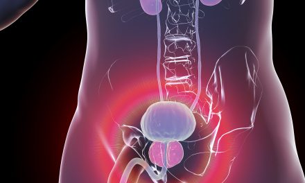
A prostate biopsy is a common procedure where small samples are taken from the prostate gland to check for cancer cells. Depending on the method, the biopsy can be done through the rectum or perineum.
These samples are then examined under a microscope to look for abnormal cells. The procedure is often guided by imaging techniques, like an ultrasound, to ensure accuracy.
Patients typically consult with their healthcare providers to understand the need for a biopsy. This procedure helps doctors determine the presence of prostate cancer, its aggressiveness, and the best treatment options.
Some of the most common methods include transrectal ultrasound-guided biopsy, which involves using a probe to direct the procedure, and MRI-targeted biopsy, which offers more precision.
The choice of biopsy type often depends on the patient’s situation and the healthcare provider’s recommendation. New techniques, like robotic-assisted biopsies, are becoming popular for their precision and reduced complication rates, offering hope for improved diagnostics.
Key Takeaways
- A prostate biopsy checks for cancer by taking tissue samples.
- Different types of biopsies are chosen based on medical advice.
- New techniques improve biopsy accuracy and outcomes.
Understanding Prostate Biopsy
A prostate biopsy involves removing small samples of tissue from the prostate gland to check for cancer cells. It is often recommended when there are signs that might indicate prostate cancer, such as elevated levels of prostate-specific antigen (PSA) or an abnormal digital rectal exam.
What is a Prostate Biopsy?
A prostate biopsy is a procedure where a doctor takes tissue samples from the prostate gland. These samples are examined under a microscope to look for cancer cells.
The biopsy is usually performed using a needle that is guided by ultrasound, either through the rectum (transrectal) or through the perineum (transperineal).
During the procedure, local anesthesia is commonly used to minimize discomfort. The use of ultrasound helps the doctor target specific areas of the prostate where cancer is suspected.
The entire process usually lasts about 10-15 minutes.
Prostate biopsies are crucial for diagnosing prostate cancer, especially if other tests like PSA screening are inconclusive. They provide detailed information about the presence of cancer and its aggressiveness, helping in planning appropriate treatment.
Reasons for a Prostate Biopsy
There are several reasons why a doctor might recommend a prostate biopsy. One of the most common reasons is elevated PSA levels.
The prostate-specific antigen is a substance produced by both cancerous and noncancerous tissue in the prostate, and high levels might suggest cancer.
Another reason is if a digital rectal exam reveals hard or irregular areas on the prostate. This can indicate the presence of a tumor.
Sometimes, imaging tests like an MRI may also suggest suspicious areas that need further investigation.
A biopsy can confirm whether these suspicions are justified and provide crucial information about the type and grade of cancer present, if any. This helps doctors make informed decisions about potential treatments and management strategies.
Before the Procedure
Undergoing a prostate biopsy requires important steps to prepare ahead of time. Patients work closely with healthcare providers to ensure the best possible outcome, focusing on consultations and thorough preparation.
Initial Consultation and Screening
Before scheduling a prostate biopsy, the patient typically meets with a healthcare provider. This consultation often includes a discussion of symptoms and medical history.
Key screenings might include a prostate-specific antigen (PSA) blood test and a digital rectal exam. These help the urologist assess the need for a biopsy. Sometimes, an MRI is performed to provide more detailed images of the prostate.
The healthcare provider also discusses risks and benefits of the procedure. This helps the patient make an informed decision.
It’s essential that all questions and concerns are addressed during this meeting.
Preparing for the Biopsy
Preparation for the procedure involves several important steps. Patients are generally advised to stop taking blood thinners and nonsteroidal anti-inflammatory drugs a few days before the biopsy.
This reduces the risk of bleeding. It’s crucial to follow the doctor’s specific instructions.
An antibiotic may be prescribed to prevent infection. Additionally, the healthcare provider might recommend an enema to clear the bowel.
On the day of the biopsy, anesthesia options are discussed to ensure comfort. It’s important for the patient to understand each step clearly.
Following these preparations helps the procedure go smoothly and reduces potential complications.
Types of Prostate Biopsy
Prostate biopsies are crucial for diagnosing prostate cancer. There are various methods for this procedure, each with its own techniques and tools. These methods aim to improve accuracy and reduce complications.
Transrectal Ultrasound-Guided Biopsy
The transrectal ultrasound-guided biopsy is a common method used to collect prostate tissue samples. An ultrasound probe is inserted into the rectum to provide live images of the prostate.
These images guide the needle to the precise location for sampling.
Local anesthetic is often applied to minimize discomfort. Generally, multiple core samples are taken to ensure accurate analysis.
While this method is widely used, it has some drawbacks, like potential infection risks. It remains a standard procedure due to its effectiveness and accessibility.
Transperineal Biopsy
Transperineal biopsy involves accessing the prostate through the perineum, the skin between the anus and scrotum. This method is considered safer concerning infection risks compared to the transrectal approach.
Local anesthetic is generally used to make the procedure more comfortable. A grid or template is sometimes employed, which helps guide the needle accurately.
This method allows for better sampling of all prostate areas, especially anterior regions that may be harder to reach through other methods.
MRI-Guided Biopsy
An MRI-guided biopsy uses magnetic resonance imaging to provide detailed views of the prostate. This method is highly precise, especially in identifying suspicious areas that may not be visible through ultrasound alone.
The patient typically enters the MRI machine after some local anesthetic is administered for comfort.
By combining MRI with biopsy, doctors can target specific regions more accurately.
This method often results in better detection rates, especially for clinically significant cancer, but might be less accessible due to the equipment required.
The Biopsy Procedure
This section covers the steps involved in a prostate biopsy, including what happens during and after the procedure. The focus is on how the biopsy is performed, and what care is needed post-procedure.
Performing the Biopsy
A prostate biopsy involves removing small samples of prostate tissue to check for abnormal cells. The procedure begins with the patient lying on their side or back.
A PSA test is often done beforehand to indicate the likelihood of prostate cancer. Local anesthesia is used to numb the area and minimize discomfort.
A transrectal ultrasound (TRUS) guides a needle through the rectum to the prostate. This allows for precise sampling.
In most cases, 12 core samples are taken to thoroughly examine the prostate tissue for signs of cancer.
Post-Procedure Care
After the biopsy, patients may experience rectal bleeding and should rest to promote healing. Drinking plenty of fluids can help flush the urinary tract, reducing the risk of infection.
Mild pain or discomfort is common and can be managed with over-the-counter pain relievers.
It’s important to monitor for signs of a urinary tract infection, such as fever or persistent pain. Patients are usually advised to avoid heavy lifting and strenuous activities for a few days, ensuring proper recovery.
Understanding Results and Follow-Up
Understanding the results from a prostate biopsy is crucial in determining the next steps for a patient. This involves analyzing biopsy tissue, receiving and interpreting the pathology report, and discussing possible treatment options based on the findings.
Analyzing Biopsy Tissue
When a prostate biopsy is performed, the tissue samples are sent to a pathologist. The pathologist examines these samples under a microscope to identify any abnormal cells.
They look for signs of cancer, including the presence of prostatic intraepithelial neoplasia (PIN), which can indicate a higher risk of cancer developing in the future.
A key part of the analysis is the Gleason score. This system grades the cancer cells based on how similar they look to healthy cells.
A higher Gleason score suggests more aggressive cancer. The staging of the cancer, which indicates how far it has spread, might also be determined during this process.
Receiving the Pathology Report
Once the analysis is complete, a pathology report is generated. This document outlines the findings from the biopsy, including whether cancer or other abnormalities like PIN were found.
The report details the Gleason score, staging, and other relevant information about the cancer diagnosis.
Patients usually receive this report during a follow-up appointment. It’s vital for them to ask questions and understand the implications of the results.
Clear communication with healthcare providers ensures the patient knows what the findings mean and what steps may be necessary. The pathology report can be overwhelming, so taking time to understand it is important.
Discussing Treatment Options
After reviewing the pathology report, patients must discuss treatment options with their healthcare team. Choices vary depending on the cancer’s stage and aggressiveness, as highlighted by the Gleason score.
Options may include active surveillance, surgery, radiation, or hormone therapy. Each treatment has its own benefits and risks.
Active surveillance might be suitable for less aggressive cancers, while surgery might be necessary for more advanced stages.
Patients should discuss all possible treatments and side effects with their doctors to make informed decisions. Engaging family or a support system can also help in making these challenging choices.
Risks and Complications
Prostate biopsy, like any medical procedure, carries certain risks and complications. One of the most common risks is bleeding. Patients might experience some rectal bleeding after the biopsy. This usually resolves on its own.
Infections are another possible complication. The procedure can introduce bacteria into the bloodstream, leading to a potential urinary tract infection (UTI). Symptoms include fever, chills, or a burning sensation while urinating.
Patients taking anticoagulants or blood thinners have an increased risk of bleeding. It is crucial for individuals to inform their doctor about any such medications before the procedure. Adjustments may need to be made to minimize bleeding risks.
In some cases, severe infections can occur. These are less common but may require hospital treatment with antibiotics. Patients should seek immediate medical attention if they experience symptoms of a severe infection.
Overall, while side effects can occur, they are typically manageable with proper medical care and guidance from healthcare professionals.
Advancements in Prostate Biopsy Techniques
Recent advancements in prostate biopsy techniques have improved accuracy and patient outcomes.
Traditional biopsies were often blind, meaning doctors had to rely on feel rather than visual guidance. Today, many prostate biopsies use ultrasound imaging, which helps in guiding the needle with more precision.
Magnetic Resonance Imaging (MRI) plays a significant role in modern biopsies. It provides detailed images of the prostate, allowing for targeted biopsies.
According to research, the use of MRI-guided biopsies helps in better identifying suspicious areas.
Combining MRI and ultrasound can enhance biopsy accuracy. This method, known as MRI-ultrasound fusion, merges images from both tools.
It has become more common because of its effectiveness in detecting prostate cancer.
Another innovation is the development of transperineal biopsy. Unlike the traditional transrectal approach, this technique reduces infection risks by taking samples through the skin between the anus and the scrotum.
Studies like those from recent advancements indicate improvements in patient safety and diagnostic accuracy.
These advancements not only provide clearer images but also give doctors better tools to make informed decisions.
By using advanced imaging scans like MRI and ultrasound, healthcare providers can offer more precise and safer prostate cancer diagnostics.
Frequently Asked Questions
Prostate biopsy processes consider various factors such as potential long-term side effects, pain management, and recovery time.
Various methods and practices ensure patient comfort and accurate results.
What are the potential long-term side effects of a prostate biopsy?
Prostate biopsies can lead to side effects like infection, bleeding, and urinary issues. These may last for a short period, although some rare cases could extend for a longer duration.
It is important to consult with a doctor if prolonged symptoms occur.
What is the likelihood of cancer being detected in a prostate biopsy?
The likelihood of detecting cancer in a prostate biopsy depends on several factors, such as age and PSA levels.
Studies, like one from the ProtecT study, evaluate these chances, helping to predict outcomes based on initial results.
How is the pain managed during a prostate biopsy procedure?
Pain during a prostate biopsy is commonly managed using local anesthesia. This reduces discomfort and helps patients tolerate the procedure better.
Proper communication between the patient and healthcare provider is crucial for effective pain management.
Are there any new methods for conducting a prostate biopsy?
Newer methods aim to improve accuracy and safety in prostate biopsies.
Research is ongoing to refine techniques and incorporate imaging tools like MRI for guided biopsies. These advances seek to minimize invasiveness and improve diagnostic accuracy.
How long does the recovery period last after a prostate biopsy?
Recovery after a prostate biopsy usually takes a few days. Patients might experience mild discomfort and are advised to avoid strenuous activities.
It is important to follow the healthcare provider’s guidance to ensure smooth recovery.
Is anesthesia typically used during a prostate biopsy?
Yes, anesthesia is typically used during a prostate biopsy.
Local anesthesia helps numb the area, minimizing discomfort. Some situations might require further sedation, but this depends on the individual’s needs and the healthcare provider’s assessment.


