
Dental X-rays play a crucial role in maintaining oral health by helping dentists diagnose and plan treatments effectively.
Despite their benefits, many people are concerned about the safety of these procedures.
Dental X-rays are generally considered safe due to the low levels of radiation used and the adherence to strict safety guidelines.

Technological advancements in dental imaging have significantly reduced radiation exposure, making modern dental X-rays safer than those used in the past.
They offer essential diagnostic benefits that support comprehensive dental care, allowing for the early detection of issues that might not be visible to the naked eye.
X-rays are vital in treatment planning, providing detailed images of teeth and surrounding structures.
Although they involve some radiation, the risk is minimal when guidelines are followed, ensuring they contribute positively to dental health.
Key Takeaways
- Dental X-rays are safe with low radiation.
- They are essential for diagnosing dental issues.
- Modern technology has improved safety standards.
Understanding Dental X-Rays

Dental X-rays are essential tools in modern dentistry, providing detailed images that help diagnose issues not visible during a routine exam.
They come in various types, each serving different purposes and using distinct techniques.
Types of Dental X-Rays
Intraoral X-Rays are the most common. Used to detect cavities, they capture detailed images of the teeth. This category includes bitewing X-rays, which show the upper and lower back teeth and how they touch.
Periapical X-rays focus on just one or two teeth, revealing the entire tooth from crown to root. Occlusal X-rays capture the roof or floor of the mouth.
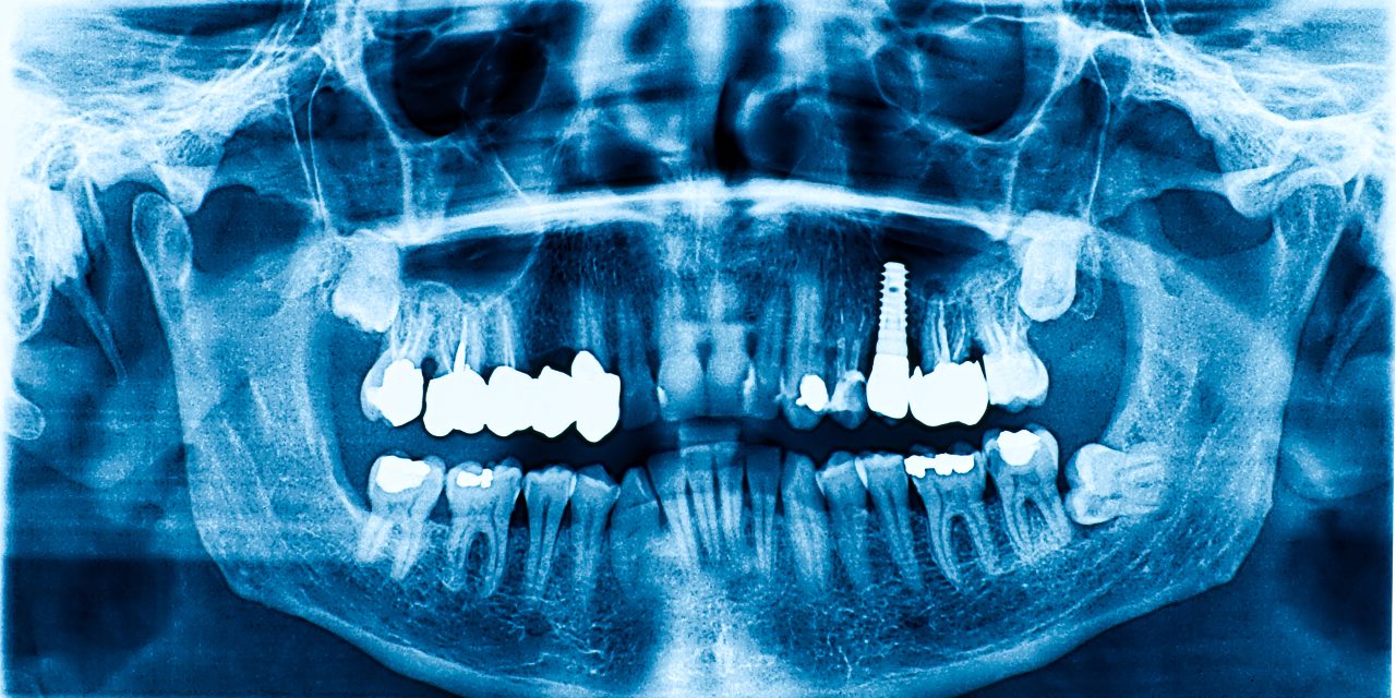
Extraoral X-Rays are taken with the film outside the mouth. They are less detailed regarding teeth but provide valuable context for jaw and skull. Panoramic X-rays offer a broad view of the mouth, showing all teeth and potential growths. Cephalometric X-rays are used to view the side of the head, useful in orthodontic planning.
Digital X-Rays are growing in popularity due to their efficiency and lower radiation levels. They offer instant images and can be easily shared between professionals.
How Dental X-Rays Work
Dental X-rays function by directing controlled amounts of radiation to create images of teeth and the surrounding jaw structure.
When a dental X-ray is taken, X-ray film or a digital sensor is placed in the mouth. The machine sends a beam through the teeth, capturing hidden issues like decay, bone loss, or abscesses.
Digital X-rays enhance traditional methods by reducing exposure to radiation and providing immediate images.
Dentists use these to evaluate treatment outcomes or monitor existing conditions.
The choice between film and digital often depends on the dental office’s resources and the patient’s specific needs.
Advances in technology continue to improve the safety and clarity of these essential diagnostic images.
Safety and Risks of Dental Radiographs

Dental radiographs are a common diagnostic tool used by dentists to assess oral health.
While they provide valuable information, understanding the levels of exposure, safety principles, and protective measures used in dental imaging is crucial to ensure safety.
Radiation Exposure Levels
Dental X-rays involve exposure to ionizing radiation, which can pose a small risk. However, the exposure levels are relatively low.
For instance, a single dental X-ray might expose a person to about 0.005 millisieverts (mSv) of radiation. To put this into perspective, this level is much lower than the average annual exposure from natural radiation sources, such as radon gas.
Safety guidelines ensure that dental radiographs are used judiciously. The risk of developing cancer from such low doses is considered minimal, especially compared to the diagnostic benefits they provide.
Dentists carefully evaluate the necessity of each X-ray to minimize unnecessary exposure.
Minimizing Radiation: The ALARA Principle
The ALARA principle, which stands for “As Low As Reasonably Achievable,” is a key guideline in radiation safety.
This principle urges dental professionals to use the lowest exposure necessary to achieve the required diagnostic results. Techniques like using digital X-rays can significantly reduce exposure compared to traditional film methods.
The ALARA principle emphasizes constant monitoring and assessment of exposure protocols.
By applying this standard, dental practices aim to protect patients while ensuring the efficacy of their diagnostic tools. Training for dental staff includes guidance on optimizing exposure settings and implementing best practices.
Protective Measures in Dental Imaging
To protect patients from unnecessary radiation, dentists commonly use protective equipment like the lead apron and thyroid collar. These devices are designed to shield vulnerable areas of the body from scattered radiation. The lead apron protects the chest and abdomen, while the thyroid collar offers additional protection for the thyroid gland.
Beyond equipment, maintaining up-to-date X-ray machines ensures that radiation output is kept within safe limits. Regular checks and compliance with safety standards are essential.
Understanding and applying these protective measures in dental imaging help minimize potential risks while maximizing the diagnostic benefits for patients.
Diagnostic Benefits in Oral Health Care

Dental X-rays are key in diagnosing oral health issues and planning effective treatments. They help in revealing diseases or conditions that are not visible during a regular dental exam. Below, the specific roles in dental health care are explored to show why X-rays are an indispensable tool.
Identifying Oral Diseases and Conditions
Dental X-rays are crucial for spotting hidden problems. They can detect cavities, gum disease, and even abscesses.
Cavities between teeth and early signs of decay are often invisible to the naked eye but appear clearly on X-rays.
In diagnosing periodontal disease, X-rays show bone loss around teeth. This helps dentists assess the severity and decide on appropriate treatments.
Identifying such conditions early prevents more complex dental issues in the future.
Role in Preventive Dentistry
X-rays play a major role in preventive dentistry by finding problem areas before symptoms develop.
Detecting dental caries in their early stages allows for less invasive treatments. This not only preserves tooth structure but also saves the patient from unnecessary discomfort.
Regular X-ray screenings help in monitoring the dental health of patients, especially those prone to tooth decay and gum disease. This proactive approach keeps teeth and gums healthy, reducing the chance of needing extensive treatments later.
Assisting Dental Procedures
X-rays assist significantly during various dental procedures. They provide a comprehensive view of the tooth structure that aids in treatment planning.
For instance, during orthodontic procedures, X-rays help in determining the position of teeth and roots.
They also guide dentists when treating root canals and in planning for tooth extractions. Clear images help ensure that procedures go smoothly with minimal risk of complications.
X-rays also aid in placing dental implants accurately, which is vital for the implant’s success and longevity.
Advancements in Dental Imaging

Recent innovations in dental imaging focus on improving precision and safety for patients. Key areas include digital imaging technologies and 3D imaging, such as Cone Beam Computed Tomography (CBCT), which offer detailed views of oral structures.
Digital Imaging Technologies
Digital imaging in dentistry has transformed the way dental professionals diagnose and treat patients.
Digital X-rays have become a popular choice due to their ability to reduce radiation exposure while providing clear images.
Compared to traditional film X-rays, digital X-rays offer quicker processing times and easier image storage. They allow dentists to zoom in on specific areas and adjust contrast to improve diagnostic accuracy. This method also supports enhanced patient communication, as practitioners can show patients real-time images during consultations.
3D Imaging: Cone Beam CT
Cone Beam Computed Tomography (CBCT) represents a significant leap in dental imaging.
CBCT provides three-dimensional images of the teeth, soft tissues, nerve paths, and bone in a single scan. Unlike regular CT scans, CBCT uses a cone-shaped X-ray beam, minimizing radiation exposure.
This technology is particularly valuable for planning complex dental procedures such as implants, orthodontics, and root canals.
Dentists can evaluate the patient’s anatomy with high precision, reducing the risk of complications.
For those interested in the technical details, CBCT plays a crucial role in modern diagnostic and treatment planning, making dental care more efficient and accurate.
Evaluation and Interpretation of X-Rays
Dental X-rays are vital for assessing dental health. These images help in identifying issues like cavities, infections, and bone loss while guiding treatment plans effectively.
Reading Dental Radiographic Examinations
Interpreting dental radiographic examinations requires knowledge and precision.
Dentists evaluate these images to detect hidden problems such as impacted teeth and potential tumors. They focus on several aspects, including the alignment of the teeth and jawbone structure.
The radiographs provide details that are not visible during a regular dental check-up. Professionals assess images for signs of tooth decay, unusual structures, and any changes in bone density.
This evaluation helps in planning treatments like root canal therapy and determining if surgical intervention is necessary.
Common Findings on X-Rays
Regular findings on X-rays may include cavities, bone loss, and signs of developmental abnormalities. Dentists often identify impacted teeth, which are teeth that have not fully emerged.
Early detection of such issues is crucial for timely treatment.
Bone loss around teeth can indicate periodontal disease. By identifying this through X-rays, dentists can take measures to address the problem and prevent further damage.
Radiographs are also used to monitor the success of procedures by observing how the teeth and supporting structures adapt over time.
Importance of X-Rays in Treatment Planning
X-rays play a crucial role in developing effective dental care plans. They help dentists in diagnosing various dental issues and deciding on suitable treatments, such as braces or root canal therapy. These images allow for precise planning of special procedures, ensuring successful outcomes.
Creating Comprehensive Dental Care Plans
X-rays provide essential information for creating a thorough treatment plan.
By revealing the condition of teeth, roots, and bone structure, they help in diagnosing dental problems that are not visible during a regular exam.
For patients requiring orthodontics, X-rays assist in assessing jaw alignment and planning the movement of teeth. They are also important in monitoring changes over time, allowing for adjustments in the care plan. This leads to tailored solutions that focus on the individual needs of each patient.
X-rays enable dentists to detect issues such as tooth decay, bone loss, and infections early. These insights help in formulating a proactive approach to prevent further complications.
Dentists can then propose preventive measures or interventions that align with the specific needs of the patient.
X-Rays for Special Dental Procedures
In cases of complex dental procedures, X-rays become even more vital.
For root canal therapy, they provide a clear view of the canal systems, assisting dentists in removing infected tissue accurately. This precision reduces the chance of complications and ensures effective treatment of dental problems.
When planning braces, X-rays allow dentists to understand the position and condition of unerupted or impacted teeth. This information is necessary to design a custom treatment plan that addresses both functional and aesthetic needs.
Certain issues, like bone infections or cysts, may require surgical intervention. X-rays offer a roadmap that guides the successful execution of such procedures. They equip dentists with the ability to anticipate potential challenges and plan accordingly, enhancing the chances of a successful outcome.
Guidelines and Frequency of Dental X-Rays
Dental X-rays are essential tools in identifying oral health issues, such as caries and periodontal diseases. Guidelines help ensure that these X-rays are used effectively and safely. Their frequency and timing are often based on recommendations and individual risk factors.
American Dental Association Recommendations
The American Dental Association (ADA) has established guidelines on the appropriate use and frequency of dental X-rays.
These recommendations take into consideration the patient’s age, health history, and risk of tooth decay. For instance, children may need more frequent X-rays due to higher caries risk and ongoing tooth development. Adults without any significant dental problems might have less frequent screenings.
Dentists follow these ADA guidelines to minimize exposure and ensure early detection of any potential issues.
The ADA emphasizes using the lowest radiation dose possible and utilizing proper shielding techniques. These best practices help protect patients while still providing essential diagnostic benefits.
Considering Individual Risk Factors
The frequency of dental X-rays isn’t a one-size-fits-all approach.
Individual risk factors such as personal and family oral health history, lifestyle choices, and environmental factors can influence the need for X-rays.
Patients with gum disease, for example, may require more frequent imaging.
Dentists assess each patient’s specific situation during an oral exam to decide the appropriate interval for X-rays. This personalized assessment ensures balanced benefits and risks, promoting optimal oral health while minimizing unnecessary exposure.
By tailoring the X-ray frequency, practitioners can ensure both safety and effectiveness in diagnostics.
Frequently Asked Questions
Dental X-rays are essential for diagnosing and treating oral health issues. They involve risks like radiation exposure, but the amounts are typically low and considered safe for most patients. Safety precautions are advised in certain cases, like for children and pregnant women.
What potential risks are associated with dental X-rays?
Dental X-rays expose patients to low levels of radiation, which is generally considered safe for most people. The primary concern is the cumulative effect of radiation over time, which could potentially increase the risk of cancer.
Professionals ensure that exposure is minimal and use protective measures like lead aprons.
How often is it recommended to have dental X-rays taken?
The frequency of dental X-rays depends on the individual’s oral health, age, and risk of dental disease.
For instance, adults with good oral health might need them every 1-2 years, while those with recurring issues might need them more often.
Dentists tailor this plan to each patient’s needs.
Are there any safety concerns with dental X-rays for children?
While the radiation exposure from dental X-rays is low, children are more sensitive to it compared to adults.
Dental professionals use the lowest possible exposure settings and may employ additional protective measures, like smaller lead aprons.
The benefits of diagnosing dental issues early usually outweigh the minimal risks involved.
What precautions should be taken for pregnant women needing dental X-rays?
For pregnant women, dental X-rays are generally considered safe, but caution is advised.
Dentists typically postpone non-urgent X-rays until after pregnancy. If an X-ray is necessary, protective measures, such as lead aprons, are used to shield the abdomen and thyroid from radiation.
Can patients decline dental X-rays and what are the implications?
Patients have the right to decline dental X-rays, but it’s important to understand the implications.
Without X-rays, some dental problems might remain undiagnosed and untreated, leading to more serious issues later.
Patients should discuss any concerns with their dentist to make an informed decision.
Are there non-radiographic alternatives to traditional dental X-rays?
Non-radiographic alternatives, like digital imaging and MRI, exist but may not always be suitable for dental diagnostics.
Digital sensors can reduce radiation exposure compared to traditional film X-rays while still providing high-quality images. Dentists can advise on the best option depending on the specific dental concern.